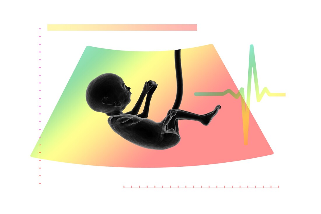
 A pregnancy ultrasound is a medical procedure that is conducted during pregnancy. It is a way to determine the number of months a woman is pregnant. Women should have their bladders full to allow a clear gel to be placed over the abdomen. This gel is applied to the pregnant woman's belly. The sonographer then moves the transducer over her belly and displays the images on a computer screen. The fetus can be heard through the monitor. A doctor will look for basic anatomy during a pregnancy ultrasound. During an ultrasound, she will look for a nuchal translucency, a fluid sack at the back of the baby's neck that contains lymphatic fluid. She will also check to see if there are any chromosomal defects or any other abnormalities. The result of the scan can be interpreted in several ways. While some problems can be diagnosed with a baby's ultrasound, others can be avoided. The first private abdominal scan is typically performed when a woman is six to eight weeks pregnant. It is also known as a baby sonogram. The cost of having a child is relatively low. However, there are a number of disadvantages to using a screening. For example, if the woman has a history of a preeclampsia, she may need to have more than one ultrasound. In addition, she may not have an accurate result if the scan is not done on her first pregnancy. During the third trimester, a woman can check the location of the placenta and see the baby. This test is called a transabdominal ultrasound. The transabdominal method is a noninvasive procedure, which can be done by either the mother or a doctor. The woman should undergo a test that identifies a uterus in the vagina. This testing is necessary for confirmation of a diagnosis of a pregnancy. A second reason for a woman to have a private ultrasound is to learn about her pregnancy risks. It can help her deliver her baby safely. It can also reveal if the fetus has certain problems that require further tests. A follow-up ultrasound can show whether the fetus has a cleft lip or is experiencing a heart defect. In some cases, a mother may need further tests besides the ultrasound to ensure that the baby is healthy. During a pregnancy ultrasound, the fetus is visible. The fetus is in the middle of the screen. The placenta is tilted towards the left of the image. The uterus is the round object at the bottom of the screen. The fetal head is positioned in the right side of the uterus. It can be seen as a mound in the center of the image. Get a general overview of the topic here: https://en.wikipedia.org/wiki/Ultrasound.
0 Comments
Leave a Reply. |
AuthorWrite something about yourself. No need to be fancy, just an overview. ArchivesCategories |
 RSS Feed
